OVERVIEW:
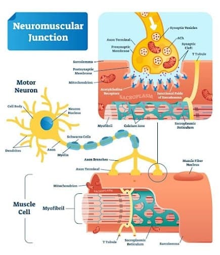
Muscles require innervation to maintain their physiological functions, like maintaining muscle tone and avoiding atrophy. A neuromuscular junction (myoneural junction) is a chemical synapse (a biological link) between a motor neuron and a muscle fibre.
Neuromuscular junction authorizes the motor neuron to carry signals from the brain to the muscles, resulting in muscle contractions.
Nerves from the central and peripheral nervous systems are interlinked and work along with muscles. Therefore, the neuromuscular junction behaves like a communication channel between the nervous system and muscle fibres.
ANATOMICAL FEATURES OF NEUROMUSCULAR JUNCTION:
The neuromuscular junction will be divided into three major parts:
- The presynaptic motor axon terminal (i.e., motor neuron).
- The synaptic cleft.
- The postsynaptic membrane (i.e., the membrane of muscle fibre cell).
PRESYNAPTIC TERMINAL:
A motor neuron consists of a dendritic end and an axonal end. Signals received from adjacent neurons are transmitted through these dendrites, whereas axons are the site from where coming signals are sent to the next neuron.
The axonal terminal of a motor neuron is termed the synaptic terminal of the neuromuscular junction. The synaptic terminal, when observed through the light microscope, consists of many tiny knobs which contain small organelles (synaptic vesicles) which are filled with neurotransmitters and are often clumped together to look like a thick membrane (dense presynaptic projection) at the terminal portion of a neuron. The presynaptic terminal is unmyelinated in structure.
ALSO, EXPLORE TRANSJUGULAR LIVER BIOPSY.
Neurotransmitters are the chemical messengers responsible for information transmission. At the neuromuscular junction, the neurotransmitter involved is Acetylcholine (Ach).
SYNAPTIC CLEFT:
It is also known as a synaptic gap of about 20 to 30 nm between the presynaptic terminal of motor neurons and the postsynaptic membrane of the muscle cell beyond which Acetylcholine (a neurotransmitter) is released. The synaptic cleft of the neuromuscular junction has a structure known as basal lamina, which possesses an enzyme (acetylcholinesterase) responsible for breaking released neurotransmitters to maintain equilibrium between their release and metabolism.
THE POSTSYNAPTIC MEMBRANE:
The postsynaptic part of the neuromuscular junction is also known as the Motor end plate. Sarcolemma (muscle cell membrane) has a thickened portion that possesses the neuromuscular junction’s postsynaptic part. It is folded in structure to form a Junctional Fold. The terminal axonal nerve ending does not perforate into the postsynaptic (i.e., motor end plate). Instead, it fits into the junctional folds.
Nicotinic Acetylcholine receptors are scattered throughout the junctional folds and have acetylcholine Ach gate ion channels. The molecule of Ach binds to these receptors and opens the channels, causing sodium influx from extracellular fluid across the motor end plate of the muscle membrane. The process results in end plate potential, producing and transmitting action potential throughout the muscle membrane.
SERIES OF EVENTS HAPPENING AT NEUROMUSCULAR JUNCTION:
The events held in the transmission of signals at the neuromuscular junction are summarised as follows:
- Acetylcholine is synthesized using choline and acetyl CoA and through the interference of the enzyme choline acetyltransferase. It undergoes a series of modulations before being packed into vesicles.
- The signal travels from the axon terminal of the previous neuron and runs down the presynaptic axonal terminal of the motor neuron, causing voltage-gated calcium channels to open. Consequently, the influx of calcium ions rushes into the nerve endings.
- An influx of calcium results in numerous neurotransmitters containing vesicles combined with the help of SNARE protein to the presynaptic neuron cell membrane.
- When they fuse with the membrane, the vesicles expel their content (i.e., Acetylcholine) outside the cell through exocytosis.
- The released Acetylcholine combines with the nicotinic ACH receptors located at the junctional folds of the motor end plate and causes an influx of sodium ions.
- End plate potential (EPP) through this sodium influx provokes an action potential that travels throughout the sarcolemma and inside the muscle fiber through T-tubules with the help of sodium-gated ion channels.
- The conduction of action potential through T-tubules triggers calcium-gated ion channels to open and release Ca++ from the sarcoplasmic reticulum, reach the myofibrils, and start contraction.
- Calcium ions can generate signals to contract other cells with the help of gap junctions, which help maintain links between the muscle cells.
Read: Unlocking the Secretes of Sciatica.
METABOLISM OF ACETYLCHOLINE (ACH):
Acetylcholine is metabolized with the help of enzymes known as Acetylcholinesterase. It is a cholinergic enzyme found in the postsynaptic membrane of neuromuscular junctions. It is responsible for the breakdown of Acetylcholine into acetate and choline. The function of AChE is to cease neuronal transmission and signaling among synapses to prevent ACh dispersal and activation of receptors.
INFLUENCE OF AGING ON NEUROMUSCULAR JUNCTION:
- With the advancement of age, there is a subsequent decline in muscle mass (sarcopenia) and weakness. These factors, along with the higher vulnerability to injury and low recovery rate, are linked to morbidity and mortality.
- In aged muscles, synaptic nuclei exhibit abnormal nuclear protein expression, like decreased levels of LMNA gene expression.
- Aging and a decrease in axonal transport are strongly associated with each other. It also disturbs the vital synaptic and energy supply to the presynaptic terminal and is responsible for age-associated changes in the neuronal cytoskeleton.
- The presynaptic structures endure degenerative changes characterized by axonal denervation, reinnervation, and remodelling concerning aging.
- The gradual changes occur at the neuromuscular junction concerning aging changes in synaptic transmission due to functional denervation in aged muscle.
- The perisynaptic Schwann cells (PSCs) are sensitive to aging; concerning age, there is shallowing of their junctional folds, which begin to retract and penetrate the synaptic cleft, which will disturb the neuromuscular junction’s structure and transmission.
- Overexpression of neurotrophins in motorneurons destabilizes NMJs by increasing agrin’s proteolytic cleavage, a signaling pathway essential for both NMJ formation.
CONCLUSION:
In conclusion, the neuromuscular junction is a remarkable and intricate interface between the nervous system and the muscular system, playing a pivotal role in the communication and coordination required for muscle contraction. Understanding the dynamics of this specialized synapse is essential for grasping the fundamentals of muscle function and the broader complexities of human physiology. With its finely tuned processes, such as neurotransmitter release, receptor activation, and signal propagation, the neuromuscular junction serves as a fundamental cornerstone of our ability to move and exert control over our bodies.
As we’ve explored in this article, the neuromuscular junction not only plays a central role in basic muscle function but also serves as a foundation for understanding various neuromuscular disorders and potential therapeutic interventions. Continued research in this field holds promise for advancing our knowledge of muscle diseases, improving our treatments, and enhancing the overall quality of life for individuals affected by these conditions.

-min.jpg)
-min.gif)
-min.jpg)
1 thought on “NEUROMUSCULAR JUNCTION: THE UNIQUE CONNECTION BETWEEN NERVE AND MYOFIBRE”